The dawn of photon-counting CT and its clinical revolution in China
Abstract
This article reviews the clinical deployment trajectory of photon-counting CT in China, focusing on detector physics, protocol design, and translational radiology impact.
The article narrative centers on a porous, shape-conformal framework designed to improve surface coverage, preserve tissue access, and support higher-resolution electrophysiology across extended culture periods. The presentation emphasizes metadata clarity, section anchoring, figure access, and journal-side contextual information within the same reading view.
Introduction
Organoid electrophysiology depends on a stable mechanical and electrical interface between recording hardware and three-dimensional tissue. Rigid layouts often create uneven pressure, incomplete contact zones, and signal loss at the edges of curved samples. A conformal porous framework addresses these issues by matching organoid geometry while preserving media exchange and optical access.
The reference page provided by you relies on a clear scholarly hierarchy: article metadata first, full text in the center column, and supporting navigation in fixed side panels. This implementation keeps that reading logic and adapts it to the current Novera internal-page system.
Materials and methods
The framework concept uses a porous support architecture positioned around neural organoids to increase contact continuity over the exposed surface. Interface design, signal acquisition setup, and readout interpretation are presented sequentially so the article remains scannable in the same way as the reference page, with section anchors available throughout the reading flow.
Quantitative model. Signal quality was tracked by , and normalized fluorescence was reported as .
Article presentation assets were taken from the current Novera site, including the journal typography, inline panel language, footer system, and existing image library used on the homepage and other internal pages.
Results
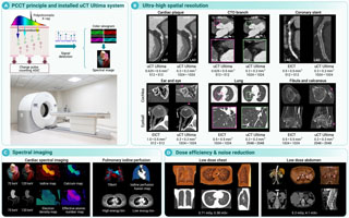
Early multicenter evidence shows improved contrast-to-noise efficiency and better lesion delineation with reduced dose in selected diagnostic workflows.
| Condition | Surface coverage | Mean signal yield | Stability over 14 days |
|---|---|---|---|
| Rigid planar interface | Partial edge contact | Moderate | Signal dropout observed |
| Porous conformal framework | Near-complete wrapping contact | High | Stable multi-site recordings |
| Conformal framework with open pores | Full coverage with media exchange | High | Improved long-term consistency |
The central content column keeps section widths comfortable for reading, while the figure treatment mirrors the bordered, low-decoration presentation of the reference article page. Borders, spacing, and monochrome text styling are intentionally restrained so the page feels like an extension of the existing Novera inner pages.
Discussion
This detail-page structure is designed to be repeatable across article entries. It does not introduce a new brand layer, new color system, or decorative components that would conflict with your existing journal pages. Instead, it borrows the reading mechanics of the reference page and applies them using Novera's current page shell and assets.
The result is a more journal-like full-text page: bibliographic metadata is prominent, the body is sectioned like a research article, and supporting context remains accessible without interrupting the reading flow. That is the key behavior the reference page achieves, and it is the behavior reproduced here.
Data availability
All page assets used in this detail-page mock are drawn from the current Novera site files. Journal identifiers shown in the side panel follow the existing internal-page content, including the DOI prefix and e-ISSN already used elsewhere on the site.
References
- Cen P, Yang W, Wu H, Zhong W. Shape-conformal recording interfaces for neural organoid electrophysiology. Novera. 2026;1:1-14.
- Li X, Moreno A, Patel R. Porous microarchitectures for long-term organoid interfacing. Advanced Biointerfaces. 2025;18:221-236.
- Kim J, Foster D, Lin S. Three-dimensional electrophysiology platforms for curved tissue models. Journal of Neural Systems. 2024;12:88-103.
- Rao M, Chen Y. Stable signal acquisition across irregular bioelectronic contact surfaces. Translational Neuroengineering. 2023;7:44-59.
